Tuesday, October 25, 2022

How X-rays are produced? __ Educational World


X-rays are commonly produced in X-rays tubes by accelerating electrons through a potential difference(a voltage drop) and directing them onto a target material(i.e. tungsten).

As these incoming electrons hit material they slow down and release X-rays(breaking radiation or bremsstrahlung). An incoming electron may also collide with an atom in the target, kicking out an electron and leaving a vacancy in one of he atom's electronic shell. Another electron may fill up this vacancy of electron and in this way an X-ray photon of specific energy is released(a characteristic of X-ray).

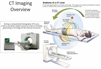
    A Computerized tomography(CT) scanner is a particular type of X-ray machine in which the X-ray tube produces a beam in the shape of a fan and moves around the patient in circle. The X-rays are detected electronically and computer uses he information to reconstruct an image of the region of body exposed.

    X-rays can also be produced by a synchrotron-a device that accelerate electron in an evacuated ring (often several tens of meters in diameter), steering them with magnet. Manipulating the electron beam in a controlled way with magnet can produce intense beam of X-rays. Synchrotron facilities are used for research purposes. 

What are X-rays __ Educational World


 X-rays are form of electromagnetic radiations, similar to visible light(400nm-900nm). In electromagnetic spectrum it lies on the left side of visible spectrum and their wavelength(0.1nm-10nm) is shorter than visible light therefore they have much energy and resolving power and are used o see small objects. As, they have higher energy they can pass through objects and therefore they have found their use in different field of life such as engineering works to check cracks in buildings and bridges.


Use in medical field to diagnose fractures

How X-rays are produced?

Computerized Tomography(CT) Scanner__ Educational World

Overview:

    A computerized tomography(CT) scanner combines a series of X-rays images taken around your body and uses computer processing to create cross-sectional images(slices) of the bones, blood vessels and soft tissues inside your body. This process is known as computerized tomography(CT) scanning and the persons that perform CT scans are called radiographer or radiology technologist. CT scan provide a clear image of body parts as compared to plain X-ray technique. For getting more clear pictures hen CT scan MRI technique is used.

    A CT Scan may have many uses but, it is particularly well-suited to quickly examine people who may have internal injuries from car accidents or other types of traumas. A CT Scan can be used to visualize nearly all parts of body and is used to diagnose disease or injury as well a to plan medical, surgical or radiation treatment.


Uses of Computerized Tomography(CT) scanner__ Educational World

 Uses:

A CT Scan help us in different ways which include:
  • Diagnose muscle and bone disorders, such as bone tumors and fractures.
  • Pinpoint the location of a tumor, infection or a blood clot.
  • Guide procedures such as surgery, biopsy and radiation therapy.
  • Detect and monitor diseases and conditions such as cancer, heart diseases, lung nodules and liver masses.
  • Used to locate infections
  • Detect osteoporosis
  • Determine the cause of chest or abdominal pain, difficulty of breathing and other symptoms.
  • Diagnose dangerous vascular diseases that can cause stroke, kidney failure and death.
  • Images the lungs to reveal blood clots in lungs vessels, excess fluid, pneumonia and chronic pulmonary obstructive disease.
  • Diagnose spinal problems and skeletal injuries.

Procedure of Computerized Tomography(CT) scanner__ Educational World


 Procedure:

    The procedure begins with lying the patient on a narrow table that slides in and out of the center of the CT scanner. The X-ray source and X-rays detectors are located on opposite sides of the scanner's ring or gantry.
During the scan, the gantry rotates around patient. The source sends X-rays through patient's body, which the detector pick up and a computer translates them into images.

    To image soft tissues of body, physician may use a special dye(called contrast dye) during the test to help visualize these body parts, which otherwise maybe difficult to see. The contrast may be ingested, given via IV, or administered rectally with an enema.

Monday, October 24, 2022

Benefits and Risks of MRI__ Educational World

 Benefits:

An MRI scanner can be used to make images of any part of body(e.g. had, joint, abdomen, legs etc.), in an imaging direction. MRI provides better soft tissue contrast than CT scan and can differentiate better between fat, water, muscle and other soft tissues than CT scan(CT is usually better at imaging bones). These images provide information to physicians and can be useful in diagnosing a wide variety of disease and conditions.

Risks:

    MRI images are made without using any ionizing radiation, so patients are not exposed to the harmful effects of ionizing radiation. But, while there are no known health hazards from temporary exposure to MRI environment, the MRI environment involves a strong, static magnetic field, a magnetic field that changes with time(pulsed gradient field), and radiofrequency energy, each of which carry specific safety concerns:
  • The strong, static magnetic field will attract magnetic objects(from small items such as keys and cell phones, to large items such as oxygen tanks and floor buffers) and may cause damage to scanner or injury, to the patient or medical professionals if those objects become projectiles. Careful screening of people and objects entering the MRI environment is critical to ensure that nothing enters the magnet area that may become a projectile.
  • The magnetic field that changes with time cause loud knocking noises which may harm hearing of patient if adequate ear protection s not used. They may also cause peripheral muscle or nerve stimulation that may feel like a twitching sensation.
  • The radiofrequency energy used during MRI scan could lead to heating of body. The potential for heating is greater during long MRI examinations.
   The use of gadolinium based contrast agents(GBCAs) also carries some risks, including side effects such as allergic reactions to the contrast agent. See GBCAs for more information.
    Some patients find the inside of scanner to be uncomfortably small and may experience claustrophobia . Imaging in an open MRI scanner may be option for some patients, bot not all MRI systems can perform all examinations.
    To produce good quality images, patients must generally remain very still throughout the entire MRI procedure. Infants, small children and other patients who are unable to lay still may needed to sedated or anesthetized for the procedure. Sedation and anesthesia carry risk not specific too MRI procedure, such as slowed or difficult breathing, and low blood pressure.

Sunday, October 23, 2022

Magnetic Resonance Imaging(MRI): Educational World

Overview:

Magnetic Resonance Imaging is a non-invasive imaging (MRI) technology that produces three dimensional detailed anatomical images. It is often used for disease detection, diagnosis, and treatment monitoring. It is based on sophisticated technology that excites and detects the change in direction of rotational axis of protons found in water that makes up living tissues.

Featured Posts

How X-rays are produced? __ Educational World

     X-rays are commonly produced in X-rays tubes by accelerating electrons through a potential difference(a voltage drop) and directing the...