Tuesday, October 25, 2022

How X-rays are produced? __ Educational World


X-rays are commonly produced in X-rays tubes by accelerating electrons through a potential difference(a voltage drop) and directing them onto a target material(i.e. tungsten).

As these incoming electrons hit material they slow down and release X-rays(breaking radiation or bremsstrahlung). An incoming electron may also collide with an atom in the target, kicking out an electron and leaving a vacancy in one of he atom's electronic shell. Another electron may fill up this vacancy of electron and in this way an X-ray photon of specific energy is released(a characteristic of X-ray).

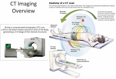
    A Computerized tomography(CT) scanner is a particular type of X-ray machine in which the X-ray tube produces a beam in the shape of a fan and moves around the patient in circle. The X-rays are detected electronically and computer uses he information to reconstruct an image of the region of body exposed.

    X-rays can also be produced by a synchrotron-a device that accelerate electron in an evacuated ring (often several tens of meters in diameter), steering them with magnet. Manipulating the electron beam in a controlled way with magnet can produce intense beam of X-rays. Synchrotron facilities are used for research purposes. 

What are X-rays __ Educational World


 X-rays are form of electromagnetic radiations, similar to visible light(400nm-900nm). In electromagnetic spectrum it lies on the left side of visible spectrum and their wavelength(0.1nm-10nm) is shorter than visible light therefore they have much energy and resolving power and are used o see small objects. As, they have higher energy they can pass through objects and therefore they have found their use in different field of life such as engineering works to check cracks in buildings and bridges.


Use in medical field to diagnose fractures

How X-rays are produced?

Computerized Tomography(CT) Scanner__ Educational World

Overview:

    A computerized tomography(CT) scanner combines a series of X-rays images taken around your body and uses computer processing to create cross-sectional images(slices) of the bones, blood vessels and soft tissues inside your body. This process is known as computerized tomography(CT) scanning and the persons that perform CT scans are called radiographer or radiology technologist. CT scan provide a clear image of body parts as compared to plain X-ray technique. For getting more clear pictures hen CT scan MRI technique is used.

    A CT Scan may have many uses but, it is particularly well-suited to quickly examine people who may have internal injuries from car accidents or other types of traumas. A CT Scan can be used to visualize nearly all parts of body and is used to diagnose disease or injury as well a to plan medical, surgical or radiation treatment.


Uses of Computerized Tomography(CT) scanner__ Educational World

 Uses:

A CT Scan help us in different ways which include:
  • Diagnose muscle and bone disorders, such as bone tumors and fractures.
  • Pinpoint the location of a tumor, infection or a blood clot.
  • Guide procedures such as surgery, biopsy and radiation therapy.
  • Detect and monitor diseases and conditions such as cancer, heart diseases, lung nodules and liver masses.
  • Used to locate infections
  • Detect osteoporosis
  • Determine the cause of chest or abdominal pain, difficulty of breathing and other symptoms.
  • Diagnose dangerous vascular diseases that can cause stroke, kidney failure and death.
  • Images the lungs to reveal blood clots in lungs vessels, excess fluid, pneumonia and chronic pulmonary obstructive disease.
  • Diagnose spinal problems and skeletal injuries.

Procedure of Computerized Tomography(CT) scanner__ Educational World


 Procedure:

    The procedure begins with lying the patient on a narrow table that slides in and out of the center of the CT scanner. The X-ray source and X-rays detectors are located on opposite sides of the scanner's ring or gantry.
During the scan, the gantry rotates around patient. The source sends X-rays through patient's body, which the detector pick up and a computer translates them into images.

    To image soft tissues of body, physician may use a special dye(called contrast dye) during the test to help visualize these body parts, which otherwise maybe difficult to see. The contrast may be ingested, given via IV, or administered rectally with an enema.

Monday, October 24, 2022

Benefits and Risks of MRI__ Educational World

 Benefits:

An MRI scanner can be used to make images of any part of body(e.g. had, joint, abdomen, legs etc.), in an imaging direction. MRI provides better soft tissue contrast than CT scan and can differentiate better between fat, water, muscle and other soft tissues than CT scan(CT is usually better at imaging bones). These images provide information to physicians and can be useful in diagnosing a wide variety of disease and conditions.

Risks:

    MRI images are made without using any ionizing radiation, so patients are not exposed to the harmful effects of ionizing radiation. But, while there are no known health hazards from temporary exposure to MRI environment, the MRI environment involves a strong, static magnetic field, a magnetic field that changes with time(pulsed gradient field), and radiofrequency energy, each of which carry specific safety concerns:
  • The strong, static magnetic field will attract magnetic objects(from small items such as keys and cell phones, to large items such as oxygen tanks and floor buffers) and may cause damage to scanner or injury, to the patient or medical professionals if those objects become projectiles. Careful screening of people and objects entering the MRI environment is critical to ensure that nothing enters the magnet area that may become a projectile.
  • The magnetic field that changes with time cause loud knocking noises which may harm hearing of patient if adequate ear protection s not used. They may also cause peripheral muscle or nerve stimulation that may feel like a twitching sensation.
  • The radiofrequency energy used during MRI scan could lead to heating of body. The potential for heating is greater during long MRI examinations.
   The use of gadolinium based contrast agents(GBCAs) also carries some risks, including side effects such as allergic reactions to the contrast agent. See GBCAs for more information.
    Some patients find the inside of scanner to be uncomfortably small and may experience claustrophobia . Imaging in an open MRI scanner may be option for some patients, bot not all MRI systems can perform all examinations.
    To produce good quality images, patients must generally remain very still throughout the entire MRI procedure. Infants, small children and other patients who are unable to lay still may needed to sedated or anesthetized for the procedure. Sedation and anesthesia carry risk not specific too MRI procedure, such as slowed or difficult breathing, and low blood pressure.

Sunday, October 23, 2022

Magnetic Resonance Imaging(MRI): Educational World

Overview:

Magnetic Resonance Imaging is a non-invasive imaging (MRI) technology that produces three dimensional detailed anatomical images. It is often used for disease detection, diagnosis, and treatment monitoring. It is based on sophisticated technology that excites and detects the change in direction of rotational axis of protons found in water that makes up living tissues.

How Does Magnetic Resonance Imaging (MRI) Works? : Educational World


 MRIs employ powerful magnets which produce a strong magnetic field that forces proton in the body to align with that field. When a radio frequency current is then pulsed through patient, the protons are stimulated, and spins out of equilibrium, straining against the pull of magnetic field. When the radio frequency field is turned off, the MRI sensors are able to detect the energy released as the proton realign with the magnetic field. The time it takes for the proton to align with the magnetic field, as well as the amount of energy released, changes depending on the environment and he chemical natures of molecules. Physicians are able to tell the difference between various types of tissues based on these magnetic properties.

To obtain an MRIs image patient is placed in a large magnetic box-in which magnetic field is present, and must remain still during the imaging process in order to make the image clear. Contrast agent(often containing the element Gadolinium) may be given to patient intravenously before MRI to increase the speed of alignment of proton within magnetic field. the faster the proton align the clear the image will be obtained.

For which purpose Magnetic Resonance(MRI) Imaging is used? : Educational World


 MRI scanner are particularly well suited to mage the non-bony parts or soft tissues of body. The differ from computed tomography(CT), in that they don't use the damaging ionizing radiations of X-rays. The brain, spinal cord, the nerve cells as well as muscles, ligaments and tendons are seen much more clear with MRI than with regular X-ray or CT scan; for this reason MRI is often used to image knee and shoulder injuries.


    In the brain, MRI can differentiate between white matter and grey matter and can also be used to diagnose aneurysm and tumors. Because MRI don't use x-ay or other radiations, it is the imaging modality of choice when frequent imaging is required for diagnosis and therapy, especially in the brain. However, MRI is more expensive than x-ray imaging or CT scanning.

    One kind of specialized MRI is functional Magnetic Resonance Imaging(fMRI). This is used to observe brain structure and determine which areas of the brain ''activate'' (consume more oxygen) during various cognitive tasks. It is used to advance understanding of brain organization and offer a potential new standard for assessing neurological status and neurosurgical risks. 

Tuesday, October 18, 2022

Astrocytoma-Brain cancer__ Educational World

Overview:

    Astrocytoma is a type of cancer that can form in brain or spinal cord. Astrocytoma begins in cells called astrocytes that support nerve cells.
    Astrocytoma signs and symptoms depend on location of tumor. Astrocytoma that occur in the brain can cause seizures, headaches and nausea. Astrocytoma that occur in spinal cord can cause weakness and disability in the area affected by the growing tumor.

Diagnosis:

Tests and procedures used to diagnose astrocytoma include:

  • Neurological exam:   During a neurological exam, doctor asks about signs and symptoms. Doctor may check vision, hearing, balance, co-ordination, strength and reflexes. Problem in one or more of these areas may provide clues about the part of your brain that could be affected by a brain tumor.
  • Imaging testing:   Imaging testing can help your doctor to determine the location and size of brain tumor. MRI is often used to diagnose brain tumors, and it may be used along with specialized MRI imaging, such as functional MRI, perfusion MRI and magnetic resonance spectroscopy.
        Other imaging tests may include CT and positron emission tomography(PET).
  • Removing a sample of tissue for testing(biopsy):   A biopsy can be done with needle before surgery or after surgery to remove your astrocytoma, depending on your particular situation and the location of your tumor. The sample of suspicious tissue is analyzed in a laboratory to determine the types of cells and their level of aggressiveness.
    Specialized tests of the tumor cells can tell your doctor the type of mutations the cells have acquired. This gives your doctor clue about your prognosis and may guide about your treatment options.

Treatment:

Astrocytoma treatments include:
  • Surgery to remove astrocytoma:   Your brain surgeon(neurosurgeon) will work to remove as much of the astrocytoma as possible. The goal is to remove all of the cancer, but sometimes the astrocytoma is located near sensitive brain tissues that makes it too risky. Even removing some of the cancer may reduce your signs and symptoms.
         For some people, surgery may be the only treatment needed. For others, additional treatments                 may be recommended to kill any cancer cells that might remain and reduce the risk that the                     cancer will return.
  • Radiation therapy:   Radiation therapy uses high energy beams, such as X-rays or protons to kill cancer cells. During radiation therapy, you lie on a table while a machine moves around you, directing beam to precise points in your brain.
         Radiation therapy might be recommended after surgery if your cancer was not removed                          completely or if there is an increased risk of cancer return. Radiation is often combined with                   chemotherapy for aggressive cancers. For people who cannot undergo surgery, radiation therapy             and chemotherapy may be used as primary treatment. 

Sunday, October 16, 2022

Gastrointestinal Carcinoid Tumor | Slow Growing Tumor


 Carcinoid tumors are type of slow-growing cancer that can arise in several places throughout the body. Carcinoid tumor which is one subset of tumors called neuroendocrine tumors, usually begin in digestive tract(stomach, appendix, small intestine, colon, rectum) or in the lungs.

Carcinoid tumors often don't cause signs and symptoms till late in disease. Carcinoid tumor can produce and release hormones into your body that cause signs and symptoms such as diarrhea and skin flushing. Treatment for carcinoid tumor usually include surgery and may include medications.(For all types of Cancer click here).

Symptoms:

Some carcinoid tumors don't cause any signs or symptoms. When they do occur, signs and symptoms are usually vague and depend on the location of tumor in body.

 Carcinoid tumors in lungs:

Signs and symptoms of carcinoid tumors n lungs include:
  • Chest pain
  • Diarrhea
  • Shortness of breath
  • Wheezing
  • Redness or feeling of warmth in your face and neck(skin flushing)
  • Weight gain, particularly around mid section and upper back
  • Pin or purple marks on the skin that look like stretch marks.

Carcinoid tumors in digestive tract:

Signs and symptoms of carcinoid tumors in digestive tract includes:

  • Abdominal pain
  • Diarrhea
  • Rectal bleeding
  • Rectal pain
  • Nausea, vomiting and inability to pass stool due to intestinal blockage(bowel obstruction)
  • Redness or feeling warmth in your face and skin(skin flushing)

Causes:

The causes of carcinoid tumors are not much clear. In general, cancer occurs when a cell develop mutation in its DNA. The mutation allow the cell to continue growing and dividing when healthy cells would normally die. The accumulating cells form a tumor. cancer cells can invade nearby healthy tissues and spread to other parts of body.

Doctors don't know what causes the cell mutation that can lead to carcinoid tumors. But, they know that carcinoid tumors develop n neuroendocrine cells.
Neuroendocrine cells are found in various organs throughout the body. They perform some nerve cell function and some hormone producing endocrine cell functions. Some hormones that are produced by neuroendocrine cells are histamine, insulin and serotonin.

Risk factors:

Factors that increase the risk of carcinoid tumor include:

  • Older age:     Older adults are more likely to be diagnosed with carcinoid tumor than are young people or children.

  • Sex:  Women are more likely to get suffer from carcinoid tumor then men. 

  • Family history:   A family history of multiple endocrine neoplais, type 1 (MEN 1), increases the risk of carcinoid tumors. In people with MEN 1, multiple tumors can form in glands of the endocrine system.

Complication:

The cells of carcinoid tumor can release hormone and other chemicals, causing a range of complications including:
  • Carcinoid syndrome:   Carcinoid syndrome causes redness or feeling of of warmth in your face and neck (skin flushing), chronic diarrhea, and difficult breathing, among other signs and  symptoms.
  • Carcinoid heart disease:   Carcinoid tumor may secrete hormones that can cause thickening of the lining of heart chambers, valves and blood vessels. This can lead to leaky heart valves and heart failure that may require valve replacement surgery. Carcinoid heart attack may usually be controlled with medications.
  • Cushing syndrome:   A lung carcinoid tumor can produce excess of a hormone that body to produce much more of the hormone cortisol.

Friday, October 14, 2022

Anal Cancer | AIDS related Cancer


 Anal cancer is an uncommon type of cancer that occurs in the anal canal. The anal canal is a short tube at the end of your rectum through which stool leaves your body. Anal cancer can lead to symptoms similar to rectal bleeding and anal pain.

Most people with anal cancer are treated with a combination of chemotherapy and radiation. Through combining the chances of cure of anal cancer increases to great extent, but it can also increase the risk of side effects.(For all types of cancer click here).

Symptoms:

   Anal cancer sign and symptoms include:
  • Bleeding from he anus or rectum
  • Pain in the area of anus
  • A mass or growth in the area of anal canal
  • Anal itching

Causes:

Anal cancer result from a genetic mutation, which may be due to any factor, which result in the abnormality of cells. The normal cells grow at normal rate and die at their set time. But, abnormal cells don't do so. They start to divide in an uncontrolled manner and also they don't die at set time due to which they accumulate in cells. This accumulation result in the formation of tumor. Cancer cells can invade near by tissues and can separate from initial tumor to spread elsewhere in body(metastasis).
        Anal cancer is closely related to a sexually transmitted infection called as human papillomavirus(HPV). Evidence of HPV is detected in majority of anal cancer patients. HPV is though to be most common cause of anal cancer.

Risk Factors:

Several factors have been found to increase the risk of anal cancer, including:
  • Older age:   Most cancer of anal cancer occur in people that are elder than 50 years.
  • Many sexual partners:   People who have many sexual partners over their lifetime have greater risk of anal cancer.
  • Anal sex:   People who engaged in receptive anal sex have an increased risk of anal cancer.
  • Smoking:   Smoking cigarettes may increase risk of anal cancer.
  • Human papillomavirus:   HPV infection increase risk of several cancers, including anal cancer and cervical cancer. HPV infection is sexually transmitted infection that can also cause genital warts.
  • Drugs or condition that suppresses your immune system:   People who take drugs to suppress their immune systems(immunosuppressive drugs), including people who have received organ transplant, may have an increased risk of anal cancer. HIV--the virus that causes AIDS-- suppresses the immune system and increases the risk of anal cancer.

Thursday, October 13, 2022

Primary CNS Lymphoma Cancer ---AIDS Related Cancer


 CNS lymphoma or primary CNS lymphoma is a rare cancer that affect your central nervous system(CNS). The cancer may originate from your brain, spinal cord, spinal fluid or behind your eye. It can spread throughout your central nervous system quickly. Treatment options vary according to patient condition.

When lymphoma starts in the CNS and isn't found anywhere else in your body, it is called primary CNS lymphoma. If lymphoma is found in other parts of body, as well as, your CNS, it is called secondary CNS lymphoma. Secondary lymphoma starts in white blood cells called lymphocytes that are he part of your lymphatic system. Lymphatic system is an important part of immune system. It helps body to fight against infections and diseases.(For all types of cancer click here).

Symptoms:

Symptoms of CNS lymphoma depend upon the location of tumor, a mass of cancerous cells. For instance, CNS lymphoma won't cause symptoms if he tumor is located in the membranes covering brain and spinal cord(meninges). In contrast, tumor near one or both eyes often cause vision changes. If the mass occur near the area of your brain that control movement, you could have weakness or co-ordination changes.
Symptoms of CNS lymphoma includes:
  • Nausea and vomiting.
  • Weakness in your arms, leg or face.
  • Weakness affecting on side of body.
  • Hearing loss.
  • Difficulty in swallowing.
  • Signs of brain pressure(headaches, confusion).
  • Vision problem(blurry vision, seeing double, floaters).
  • Changes in your mental state(memory loss, trouble speaking, feeling sluggish).
  • Seizures(that may become more frequent several days or weeks).
  • Trouble controlling when you pee or poop(incontinence).

How is CNS lymphoma diagnosed?

Healthcare provider may adopt different methods to diagnose CNS lymphoma. Some of them are listed below:  

Exams:

Your healthcare provider may perform different kinds of exams to diagnose CNS lymphoma.
  • Physical exam:  Your provider will assess your over health by examining your body and asking questions about your medical history, lifestyle and symptoms
  • Neurological exam:   Your provider will perform neurological exam to check how your central nervous system functions. They may check your reflexes, movement, co-ordination and ability to process information and speak. 
  • Exam eyes:  Your provider may perform various test to exam your eye to find tumors behind your eye which normally affect vision. A slit lamp examination is commonly used as a part of diagnosis of CNS lymphoma. During the procedure, your provider uses a special microscope called slit lamp to see parts of your eye magnified. They can also detect signs of tumors behind your eye, like swollen optic nerves.

Imaging Procedure:

Your healthcare provider may perform tests to see where cancer is located inside of your body. CNS lymphoma rarely spread beyond your central nervous system, but it may spread quickly through various parts of CNS.
  • MRI:   An MRI(magnetic resonance imaging) uses magnets and radio waves to scan your body and produce a detailed image of your inside body inside computer. Your provider may give you a harmless substance called gadolinium by injection to make the cancer show up better. Once it's inside of your body, Gadolinium clusters around cancer cells, making them stand out more on computer.
  • CT scan:   A CT scan(computerized tomography) uses X-ray to scan your body and produces a detailed image of the soft tissue and bones inside of your body. A CT scan may be used to show if the cancer has spread beyond your CNS or not.
  • PET scan:   During a PET scan(positron emission tomography), you will receive an injection of radioactive tracer that travels through your body and collects near diseased cells. Meanwhile, a machine takes multiple images of the inside of your body. The tracer makes the cancer cells stand out in the images. Like a CT scan, your provider may order a PET scan to track cancer spread. Sometimes, the two procedures take place together and produce a three dimensional image(3D) that makes abnormal cells easier to see. This is called a PET-CT scan.  


Wednesday, October 12, 2022

AIDS-Related Lymphoma


 AIDS related lymphoma is a disease in which cancerous cells(malignant tumors) are found in the lymph system of patients who have AIDS.

The lymph system is made up of thin tubes that branch, like blood vessels into all parts of body. Lymph vessels carry lymph, a colorless, watery fluid that contain white blood cells called lymphocytes. Along he network of vessels are group of small bean shaped-organs called lymph nodes. Cluster of lymph nodes make lymphocytes and filter old blood cells from the blood; the thymus, a small organ beneath the breast bone; and the tonsils, an organ in the throat, are part of lymph system.

Because there are lymph tissues in many parts of body, the cancer can spread to all parts of body's organs and tissues including liver and bone marrow(spongy tissue inside the large bones of body that makes blood cells), spleen or brain etc.(For all types of cancer  click here ).

Types of Lymphoma:

Lymphoma is generally categorized into two main types:

  • Hodgkin's lymphoma
  • Non-Hodgkin's lymphoma


Kaposi Sarcoma


 Kaposi sarcoma is a disease in which cancer cells are found in skin or mucous membranes that cover the wall of gastrointestinal(GI) tract from moth to anus including stomach and intestines-means the whole alimentary canal. These tumors appear as purple patches or nodules on the skin or on mucus membranes and can spread to lymph nodes and lungs. Kaposi sarcoma is more common in men and in patients with suppressed immune system.(For all types of cancer click here).

What cause Kaposi sarcoma?

Kaposi sarcoma is always caused by an infection with a virus called human herpes virus 8, which is also known as Kaposi sarcoma-associated herpes virus(KSHV). The virus, which is in the same family as Epstein-Barr virus, is rare in United States. In fact, less than one percent of general US population is carrier. The virus and tumors are much more common in some other parts of world.

How the virus is initially acquired and spread is poorly understood, but scientists have identified four distinct populations that represent near all cases of disease. There is some evidence within those populations as to how KSHV is acquired and what causes some carrier to develop Kaposi sarcoma.

What are the risk factors for Kaposi Sarcoma?

The following factors can raise a person's risk of developing Kaposi Sarcoma:
  • Ethnicity:   People of Jewish or Mediterranean descent, as well as equatorial Africans have higher risk of developing Kaposi sarcoma.
  • Sex:   Men have higher risk of developing Kaposi sarcoma than females.
  • Human herpes virus 8 (HHV-8):   This virus, also called the Kaposi sarcoma herpes virus (KSHV) can cause Kaposi sarcoma. However, most people with HHV-8 infection do not develop Kaposi sarcoma. The cancer appears most often when a person with HHV-8 also has problems with their immune system function.
  • Immune deficiency:   People with human immunodeficiency virus(HIV) and acquired immune deficiency syndrome(AIDS) and people whose immune system are suppressed after organ transplantation have a higher risk of developing Kaposi sarcoma.
  • Sexual activity:   Unprotected sexual activity can increase the risk of infection with HVV-8 as well as HIV, because these viruses are spread through body fluids such as semen, blood and sera.  

Adrenocortical Carcinoma Cancer

 Adrenocortical carcinoma ,or ACC, is a cancer of adrenal gland, which are two small triangular-shaped glands that sit on the tope of each kidney. The outside of these glands is called adrenal cortex. The adrenal cortex makes important hormones that help your body control water balance, blood pressure, stress response and cause the body to have male or female traits. ACC originate from adrenal cortex.

An ACC may be functioning, which means it makes more hormone than normal, or non-functioning, which means it has no effect on hormone production. A functioning ACC tumor makes to much of the hormone cortisol, aldosterone or estrogen(For all types of cancer click here).

How common is adrenocortical carcinoma?

ACC is very rare, affecting around one case diagnosed in one million people in the US. It is more common in females as compared to males.

Tuesday, October 11, 2022

Cancer

 What is cancer?

Definition:  

Cancer is a disease in which some of the body cells grow in uncontrolled fashion and spread to other other parts of body resulting in tumor.
Cancer can start  almost anywhere in the human body which is made of trillions of cells. Normally, the cells of human body grow and multiply, by a process called cell division, to form new cells. When the cells of body are worn out they die and are replaced by new one.
sometimes this orderly process of cell division breaks down and abnormal cells are produce. When the cycle break the cells start dividing in an uncontrolled fashion and form bulk of abnormal cells called tumor. Tumors can be cancerous or non-cancerous(benign).(For all types of cancer click here).
Cancerous tumors can spread and invade into near by body tissues and form tumors in other parts of body (a process called metastasis).Cancerous cells are also called malignant tumors. Many cancer form solid tumors but some type of tumors like leukemias(blood cancer),generally do not.
On the other hand benign tumors do not invade into other body tissues. When removed, benign tumors usually do not grow back. Benign tumors can sometimes be quite large. Some benign tumors are also life threatening, such as benign tumor in brain.

Types of Cancer(A--Z all types of cancer in detail)

 There are hundreds types of cancer. All the types are listed in alphabetical order. You can also find your related types by writing first alphabet of you related type in search engine.

Here, the list of some common types is given which may help you in finding your related types:
  • Bladder Cancer
  • Breast Cancer
  • Colon and Rectal Cancer
  • Endometrial Cancer
  • Kidney Cancer
  • Leukemia 
  • Liver Cancer
  • Lung Cancer
  • Melanoma AIDS-related lymphoma
  • Non-Hodgkin Lymphoma
  • Pancreatic Cancer
  • Prostate Cancer
  • Thyroid Cancer

A

AIDS-Related Cancers
   AIDS-related lymphoma(lymphoma)

Acute Myeloid Leukemia

 Acute myeloid leukemia(AML) is a type of blood cancer that start in bone marrow the soft inner part of bones. AML usually begins in the cells that are later transformed into white blood cells, but t can affect the other blood-forming cells, as well.

With acute form of AML, the bone marrow do not grow in the fashion in which it has to grow. These immature cells-blasts, build up in body. (For all types of cancer click here).

The other names for leukemia are:

  • Acute myelocytic leukemia
  • Acute myelogenous leukemia
  • Acute granulocytic leukemia
  • Acute non-lymphocytic leukemia
If you don't get medical treatment AML can be life threatening. It can spread quickly to your blood and to other parts of body: such as,
  • Lymph nodes 
  • Liver
  • Spleen
  • Brain and Spinal cord
  • Testicles

Leukemia-Blood Cancer

 Leukemia is the broad term for cancers of the blood. It is further subdivided into various types depending upon the types of blood cells which become cancerous. Leukemia is more common in adults older than 55, but it is also the most common cancer in children younger than 15 years.

Acute lymphocytic leukemia((ALL) is a type of blood cancer  which affect blood and bone marrow cells-the spongy tissue inside bone where blood cells are formed.(For all types of cancer click here).

The word "acute" in acute lymphocytic leukemia comes from he fact the disease progresses rapidly and creates immature blood cells, rather  than mature ones. The word "lymphocytic"  in acute lymphocytic leukemia refers to the white blood cells called lymphocytes, which ALL affect. Acute lymphocytic lekima s also known as acute lymphoblastic leukemia. 

Featured Posts

How X-rays are produced? __ Educational World

     X-rays are commonly produced in X-rays tubes by accelerating electrons through a potential difference(a voltage drop) and directing the...